HOW TO READ CHEST XRAY- Basic fundamentals
X-ray is a type of radiography and most widely used investigation. It first appears too complicated to read the chest xrays because we barely know what lies where and what to make out of it.
Here,we will be explaining step-to-step study of Chest Xrays and identify the diagnosis.
Basics of X-Rays to identify:-
1. PA (posteroanterior) or AP (anteroposterior) view
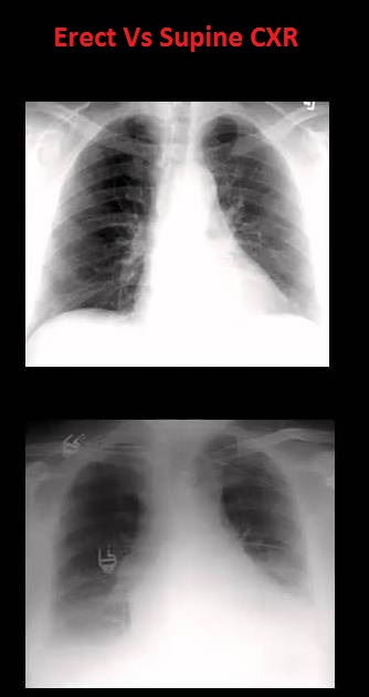
2. Erect or Supine position of patient
Let's look into them individually to find how can we tell if we see a chest X-Ray:-
AP or Anteroposterior view:
The view is from front to back
PA or Posteroanterior view:
The view is from back to front
Features
|
PA view
|
AP view
|
Position of clavicle
|
Oblique
|
Horizontal
|
Scapula
|
Away from lung field
|
Over the lung field
|
Spirolamina angle
|
Inverted ‘V’
|
Not significant
|
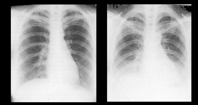
PA is most common X-Ray done where AP is usually done when patient cannot stand and XRay machine is brought to him on bed and view taken from anterior to posterior.
Cardiomegaly in Chest Xray
The point to add is that there is apparent Cardiomegaly in AP view as compared to PA view because there is slight magnification of heart since heart is away from view capturing film. This can be well understood by the following:-
The approach to cardiomegaly on Chest Xray is as follows:-
A/B x 100 = cardio ratio
In PA view, Cardiomegaly when ratio is more than 50%.
In AP view, Cardiomegaly when ratio is more than 60%.
There is fundal view in erect position because all the air in stomach comes in fundus when the patient is standing.
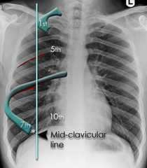
If anterior end of 6th or 7th rib reaches mid-clavicular line of diaphragm, it is Inspiratory Xray.
Counting Ribs in Chest Xray:-
Two points can just help you quickly count ribs from top to bottom:-
1. The front opaque appearing side of ribs is actually it's posterior side.
 |
| Anterior and Posterior side of ribs on chest xray |
Before we proceed, let us see what structures lie in a normal Chest Xray:-
 |
| Structures seen on normal Chest Xray |
The Chest Xray is usually divided into three zones as:-
Upto 2nd rib- First zone
 |
| Zones of chest xray |
Upto 2nd rib- First zone
2nd to 4th rib- Second zone
4th to 6th rib- Third zone
4th to 6th rib- Third zone
Now let's proceed to start studying the Xray:-
Lucency and Opacity in Chest Xray:-
Lucency-
Anything that appears dark or black on chest xray is said to be lucent.
-This is because of less density.
-Black color appears because of AIR.
-Black color appears because of AIR.
Opacity-
Anything that appears light or white on chest xray is said to be Opaque.
-This is because of high density.
-White color appears because of Bones and soft tissues.
-White color appears because of Bones and soft tissues.
Therefore, we can conclude the following easily:-
Increase in lucency:
- Increase in air
-Decrease in soft tissues or absence of bone
-Decrease in soft tissues or absence of bone
Increase in Opacity:
-Increase in soft tissue or abnormal bone
-Decrease in air
-Decrease in air
The basic approach when seeing a chest xray always sequentially as:-
1. Define whether xray is normal or abnormal.
2. If xray is abnormal, where is this abnormality.
3. Extent of abnormality
4. What is the final diagnosis
2. If xray is abnormal, where is this abnormality.
3. Extent of abnormality
4. What is the final diagnosis
Before we proceed to pathological approaches to Chest X-Rays, let's see what layers the xrays hit when they enter the body. Note this strengthens further basics:-
Muscle> Ribs> Pleura> Lung
Talking about when Hyperlucency (increase in blackness) or Hyperopacity (increase in whiteness) occurs:-
1. Consolidation- Replacement of air by something abnormal
2. Atelectasis- Collapse of lung resulting in loss of air
Also seen in Plethora, i.e, increase i vascularity.
The differential diagnosis of three important causes if unilateral (one side) opaque thorax are:-
-Displacement of interlobar fissure: because the lobes of lung collapse, the fissures in between the lobes move up or down because of hyperinflation of normal lobe against collapsed lobe.
This is the most reliable direct sign of Collapse.
This is the most reliable direct sign of Collapse.
-Mediastinal shift: The structures on mediastinum shift to side of collapsed lung.
-Crowding of ribs
-Elevation of hemidiaphragm
-Sharp defined margins of opacity.
-No mediastinal shift
-Ill defined margins of opacity
-Airbronchogram sign: visualization of air in bronchus sorrunded by alveolar opacity
*image*
Positive Airbronchogram sign is seen in:
-All except interstitial (viral) pneumonia
-Pulmonary oedema (water replace air)
-ARDS (Acute respiratory distress syndrome)
-Goodpasture syndrome (blood)
-HMD (Hyaline membrane disease)
-Pulmonary alveolar proteinosis (macrophages congested in alveoli making crazy paving pattern)
-Pulmonary oedema (water replace air)
-ARDS (Acute respiratory distress syndrome)
-Goodpasture syndrome (blood)
-HMD (Hyaline membrane disease)
-Pulmonary alveolar proteinosis (macrophages congested in alveoli making crazy paving pattern)
Airbronchogram sign is NOT seen in:
-Lung abscess
-All except bronchoalveolar carcinoma
-All except bronchoalveolar carcinoma
3. Pleural effusion-accumulation of fluid
Normally, there is no air in pleura. But effusion in pleura can occur.
-Mediastinal shift: which is on opposite side, i.e, structures shift to opposite side of pleural effusion.
Note: Pleural effusion and Haemothorax cannot be differentiated because soft tissue cannot be differentiated on Chest Xray.
-Rotation: apparent increase in air gap
-Scoliosis
-Scoliosis
-Masectomy
-Poland syndrome (absent pectoralis major muscle)
-Airway obstruction
-Large pulmonary embolus
-Pneumothorax
-Airway obstruction
-Large pulmonary embolus
-Pneumothorax
A small pneumonic to quickly grab the names:-
POEMS
POEMS
P- Poland syndrome/Pneumothorax
O- Oligemia/Obstruction (like Pulmonary embolism)
E- Emphysema
M- Mastectomy/Mucous plug
S- Swyer's james syndrome
Enjoyed reading? In our upcoming blog of Chest xray, we will be explaining Silhoutte sign (where exactly the abnormality is in the lung relating with intervening border of an organ or its part) and some pathological diseases observed on chest xrays like pulmonary embolism, left ventricular failure, bronchogenic carcinoma, bronchiectasis, and lots more. Stay tuned.


